
How Does Breast Ultrasound Imaging Work?
During your Breast Ultrasound Screening appointment with HerScan, the screening may seem quick and easy (taking approximately 15 minutes), but there is a lot going on behind the scenes. The breast imaging technology used in this type of screening is complex and offers invaluable insight into breast health, especially for women with dense breast tissue. Continue reading to learn about how breast ultrasound imaging works and what to expect during your appointment.
Breast Ultrasound Imaging
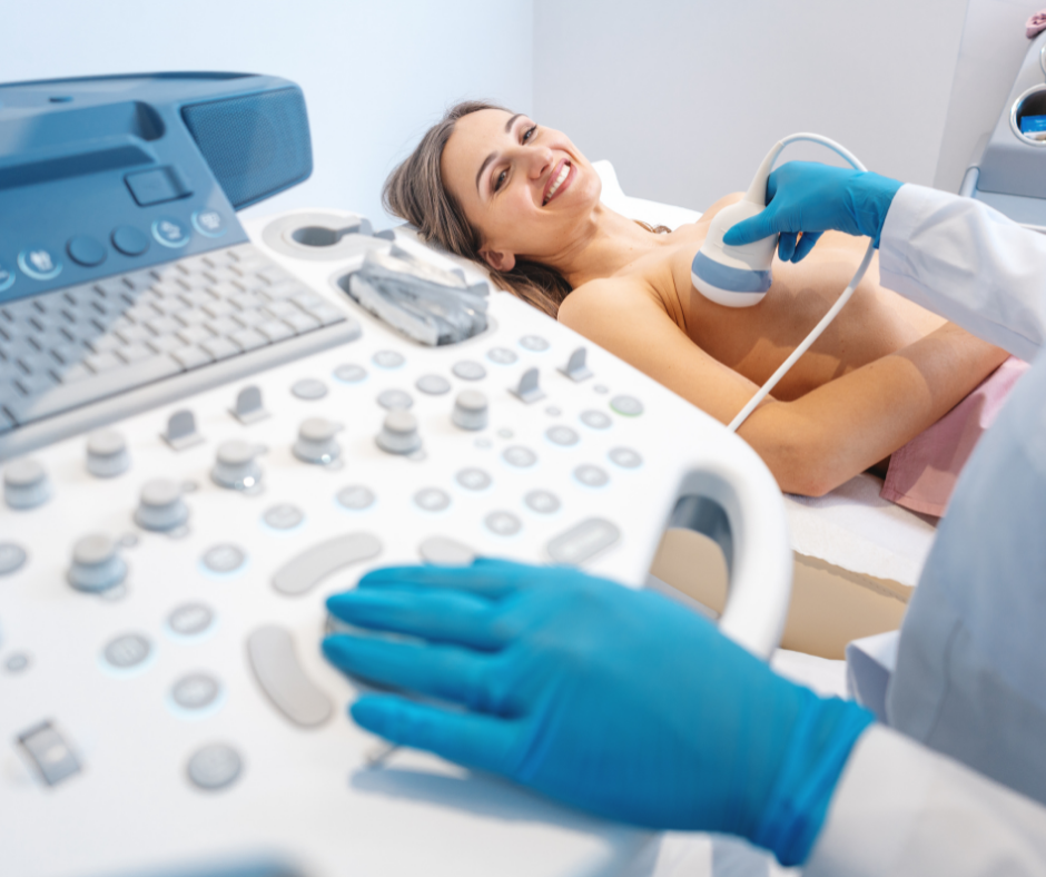
Breast ultrasound imaging uses sound waves to see inside your breasts to detect abnormalities and masses. In addition, breast ultrasound imaging also detects blood flow throughout the breasts. To see inside the body, a wand-like transducer glides over both breasts and the axilla (armpit) area in a repeated motion. A warm gel is applied to this area and then it is ready to be probed by the transducer. The transducer device is responsible for sending high-frequency sound waves through the breast tissue. The sound waves bounce back and forth through the body, creating an echo. The echo waves are then measured, producing the imaging that is viewed on a computer screen. A Radiologist interprets the internal structures of the breast and then determines the results after the screening is complete.
Since breast ultrasound imaging uses sound waves to see through the breast and does not emit any radiation, it is safe for breastfeeding mothers, pregnant women and other vulnerable populations.
Ultrasound and Breast Density
No breast screening tool is perfect; while mammography offers a range of benefits for early cancer detection, it does have limitations, especially for women who have dense breast tissue.
Breasts contain three types of tissue: glandular, connective and fat tissue. Simply put, dense breast refers to the amount of fibrous and glandular tissue in a woman’s breast compared to fatty tissue as seen on a mammogram.
Dense breast tissue is composed of more fibrous or glandular tissue than fat, making it much more difficult to interpret on a mammogram. Approximately 50% of the women in the United States have dense breast tissue. On a mammogram, dense tissue appears white, while cancerous tumors also appear white, making it nearly impossible to "see" the tumor. It's like trying to finding a snowball in a snowstorm. This is why ultrasounds are so critical for women with dense breast tissue, as nearly half of all cancers are missed with mammography alone.
Breast ultrasound is a powerful screening tool that can see beyond glandular and connective tissue. Studies have found that mammography misses every other cancer in dense breasts. Breast ultrasound has been shown to increase early detection rates of breast cancer from approximately 48% to 97% in women with dense breasts, making it an invaluable screening tool to add to your breast health regimen.
What to Expect During Your Breast Ultrasound Exam
You’ll be happy to know that breast ultrasound screening is completely painless and compression-free. Many women find it to be an easy and relaxing experience that can even be performed during their lunch break.
During the time of your appointment, you will lay down on an exam table and be asked to raise your hand over your head so the RDMS Sonographer can move the transducer device over the breast and armpit area. The transducer will glide over this area until the desired images are produced on the computer screen.
After the completion of your screening, your test will be read by our board-certified radiologist. You will receive your completed report and images within 10 business days via Our New Ramsoft Omega AI Blume Patient Portal.
Do more for your breast health. Add powerful breast Ultrasound Screening to your healthcare routine. For more information on HerScan and to request an appointment, please click here.